
Description
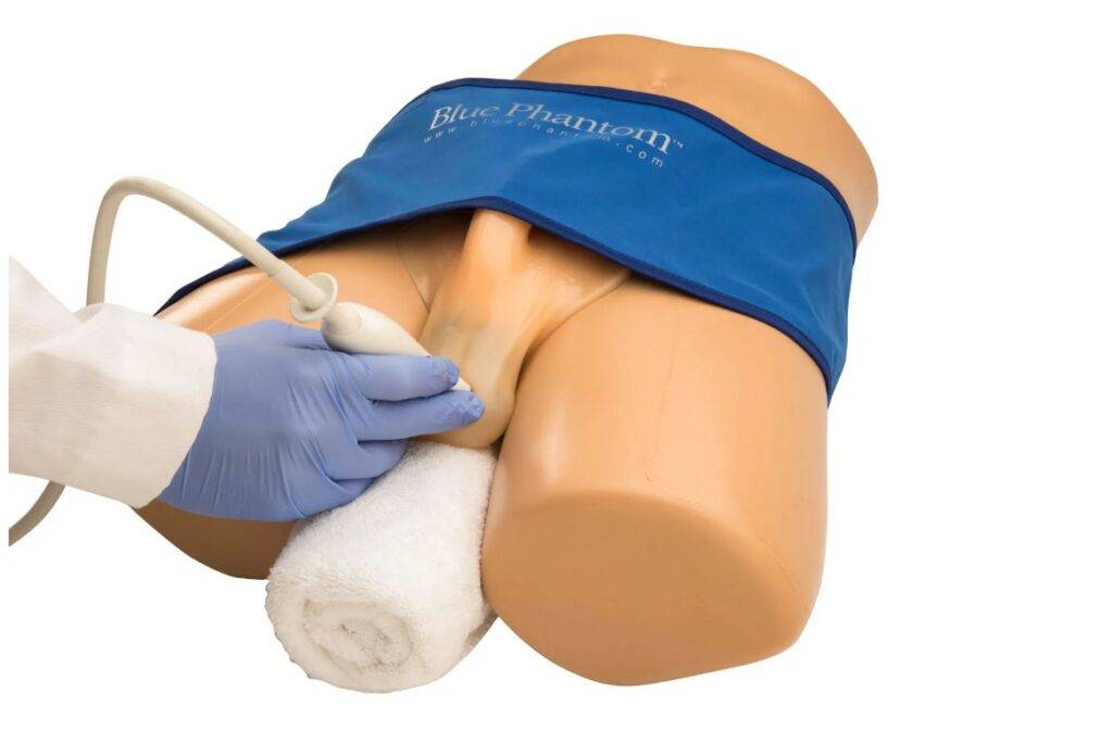
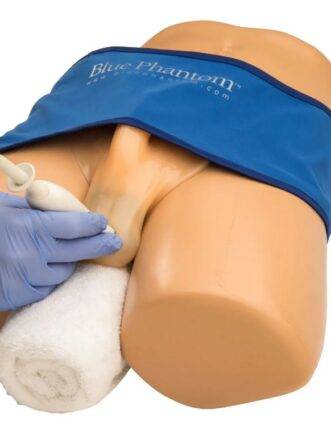
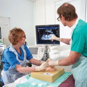
Adam,Rouilly offers a wide selection of Clinical Skills Simulators – Ultrasound – Andrology/Pelvic Examination. This Sale Price Blue Phantom Scrotal Ultrasound hands-on training model offers users an excellent training platform for teaching and learning how to perform scrotal ultrasound procedures using their own ultrasound system.
Skills
- Blue Phantom’s Scrotal Ultrasound hands-on training model offers users an excellent training platform for teaching and learning how to perform scrotal ultrasound procedures using their own ultrasound system
- Excellent for training users to develop and practice patient setup, image acquisition, interpretation, and the psychomotor skills necessary to perform scrotal ultrasound exams
- Gain proficiency in performing scrotal ultrasonography including patient setup and identification of normal scrotal structures and pathology
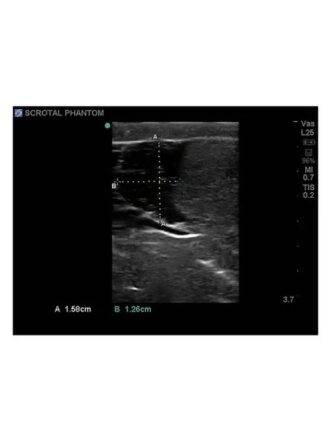
- Develop, practice, and verify user’s ultrasound imaging skills including; using ultrasound system controls, transducer positioning and movement, recognition of scrotal anatomy and pathology, and use of ultrasound calculation packages to measure cysts, masses, and other structures
- Tissues match the acoustic characteristics of real human tissue so when you use your ultrasound system on our training models, you experience the same quality you expect from imaging patients in a clinical environment
- Practice using ultrasound system controls
- This ultrasound phantom model is excellent for specialties including urology, emergency medicine, fertility training programs, ultrasound training programs, simulation centres, medical education facilities, and ultrasound manufacturers for ultrasound education and demonstrations
Features
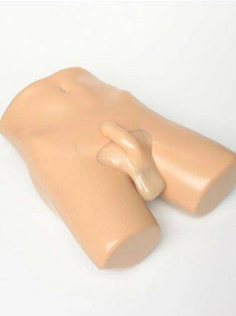
- Blue Phantom’s scrotal ultrasound training models contain the external anatomy of the male lower pelvis, penis and scrotum
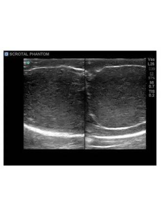
- Model contains anatomically correct internal and external anatomy with both normal and pathological scrotal structures represented
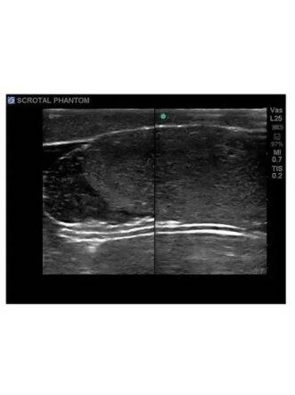
- Contains a normal testis and epididymis and contralateral testis and epididymis with pathology to be used for comparison
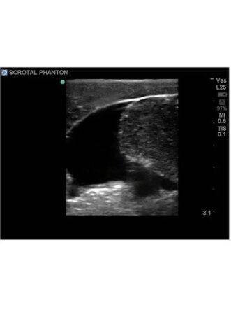
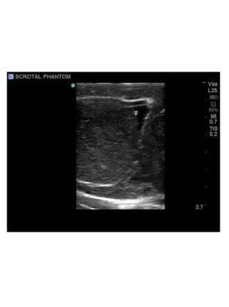
- The contents of the scrotum are fully imageable using real-time 2-D ultrasound imaging and contain the left and right testicles with epididymal head, body, and tail as well as pathological scenarios including intratesticular masses, an epididymal mass and a hydrocele.
- This medical training model will perform well using any ultrasound imaging system configured with the appropriate scrotal ultrasound transducer
- Develop, practice, and verify user’s ultrasound imaging skills including
- Tissues match the acoustic characteristics of real human tissue so when you use your ultrasound system on our training models, you experience the same quality you expect from imaging patients in a clinical environment
- Performs well using any ultrasound imaging system
- Superb ultrasonography imaging characteristics
- Product dimensions:
- L 44cm W 33cm H 18cm
- Weight 10 Kg
Why Choose the Scrotal Ultrasound Training Model for Your Ultrasound Training Programme in India?
The Scrotal Ultrasound Training Model (Item No. ABP092) by Adam, Rouilly Limited, UK is a 10 kg Blue Phantom hands-on training platform that lets trainees perform real scrotal ultrasound scans with their own ultrasound system. For urology training centres, emergency medicine residency programmes, fertility/andrology training units, and ultrasound training institutes in India that need to teach scrotal sonography without relying on live patients, this model provides a realistic, repeatable platform for both normal and pathological findings.
The model contains the external anatomy of the male lower pelvis, penis, and scrotum, with anatomically correct internal and external structures. It includes a normal testis and epididymis alongside a contralateral testis and epididymis with pathology, allowing trainees to directly compare healthy and abnormal findings side by side during the same exam. Imageable structures and pathological scenarios include the left and right testicles with epididymal head, body, and tail, intratesticular masses, an epididymal mass, and a hydrocele — giving trainees exposure to the most common findings they will encounter in clinical scrotal ultrasound. The tissue is engineered to match the acoustic characteristics of real human tissue, so trainees see the same image quality on this model as they would scanning an actual patient. The model performs well with any ultrasound imaging system fitted with an appropriate scrotal transducer, and supports practice with transducer positioning and movement, system controls, anatomy/pathology recognition, and use of calculation packages to measure cysts and masses. Product dimensions: L 44 cm x W 33 cm x H 18 cm; weight 10 kg.
Why Buy from SEM Trainers?
SEM Trainers & Systems is an authorized distributor of Adam, Rouilly products in India with over 30 years of experience supplying medical simulators to 1,500+ institutions across 28 states. We provide pan-India delivery, on-site installation, faculty orientation, and dedicated after-sales support.
Q: Does this model work with my existing ultrasound machine?
A: Yes. The model performs well using any ultrasound imaging system configured with the appropriate scrotal ultrasound transducer.
Q: What pathologies can be demonstrated?
A: Intratesticular masses, an epididymal mass, and a hydrocele, alongside a normal testis and epididymis for comparison.
Q: What are the dimensions and weight?
A: L 44 cm x W 33 cm x H 18 cm, weighing 10 kg.
Reviews
There are no reviews yet.