Description
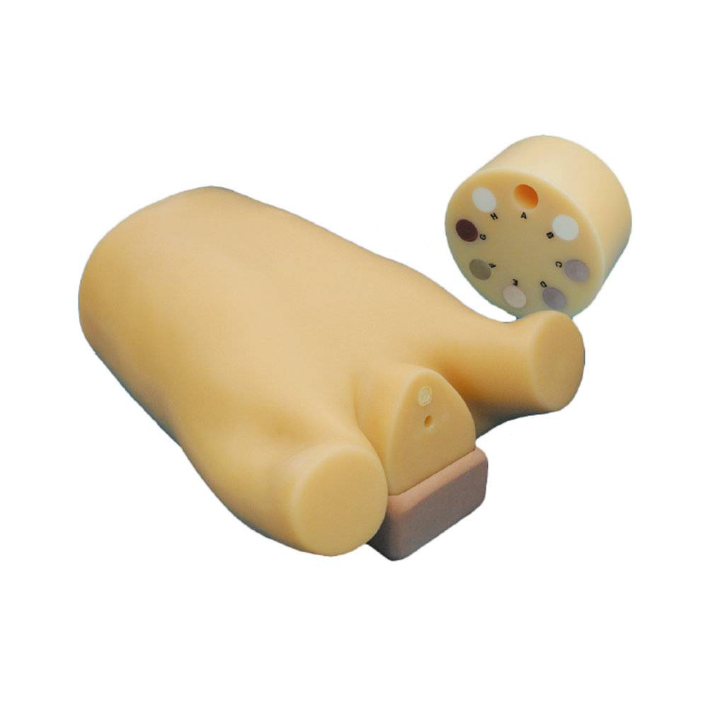
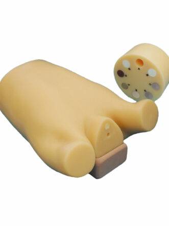
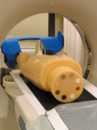
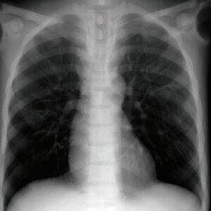
Chest phantom for standardization studies in low dose lung cancer CT screening / Anthropomorphic structure provides life-like images allowing operators visual evaluation
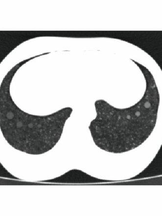
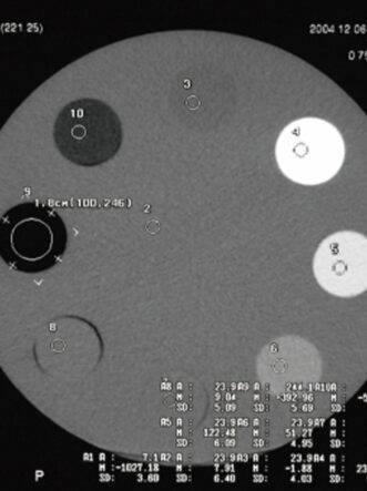
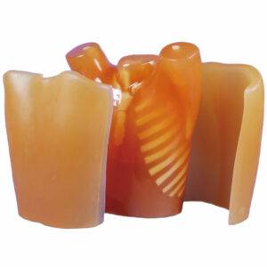
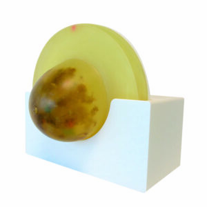
| Features | 1. Simulated GGO type tumors with different sizes and HU numbers are prepared in the vicinity of three main sections of bilateral lungs 2. Dosimeter holder on the central axis of the phantom allows housing a pencil type ion chamber. 8 step cylindrical linearity phantom to control density curve as a scale can be attached to the chest phantom base. |
|---|---|
| Set includes | 1 chest phantom / 1 8 step linearity phantom / 2 urethane cylinder / 1 adjustment base / 1 set of sample images / manual |
| Size (approx.) | chest girth 93cm H 45cm |
| Packing size (approx.) | W63 x D50 x H29cm / W24.8 x D19.7 x H11.4in |
| Weight (approx.) | 18kg |
| Materials | Chest wall: human tissue substitute / Bones: synthetic bones / Alveoli: styrene foam and urethane foam |
| Optional parts | 41363-020 Carrying case for PH-1 |
| Publication references | Muramatsu, Y., Tsuda, Y., Nakamura, Y., Kubo, M., Takayama, T., & Hanai, K. (2003). The Development and Use of a Chest Phantom for Optimizing Scanning Techniques on a Variety of Low-Dose Helical Computed Tomography Devices. Journal of Computer Assisted Tomography, 27(3), 364-374. doi:10.1097/00004728-200305000-00012 |
| ANATOMY | Bones / Lungs / Mediastinum / Simulated tumors at three lung areas / Apical portion of the lungs / Bifurcation of the trachea / Base of lungs |
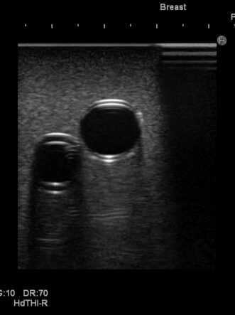
| Application | CT image quality evaluation / Dosimetry / Evaluation of density curve |
| Update | July 10, 2020 |
Reviews
There are no reviews yet.