
Description
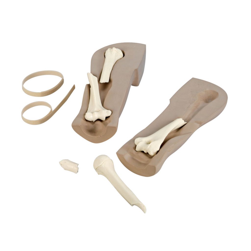
An exciting training device designed to aid both radiology educators, as well as radiology students. Educators will be able to enhance hands-on training of identifying radiographic pathology with a tool that will encourage students to utilize their critical thinking skills. Students will be excited to image a body part that will challenge their ability to create optimal images of a bone that demonstrates pathology common to the upper extremity. This life-like upper extremity is uniquely designed to permit the user access to the fracture site in which various fracture types can be easily interchanged. Student can quickly visualize the difference between a spiral fracture and a splinter fracture within minutes of taking the radiographic exposures. The simulator is designed to allow students the capability of rotating the arm easily to place the humerus in routine and special projections common to imaging with protocols.
With the Life/form® Imaging Fracture Simulator, students will be able to practice positioning skills, technical skills, and critical thinking skills as they produce radiographic images that will make learning anatomy, pathology, and image production an exciting educational experience. Exposure techniques for the simulator are in line with techniques typical to an adult humerus. Students will be able to visualize, create, and reduce bone (anterior/posterior) displacement. Imaging concepts will become easy to understand as students can see how changing the position and/or projection of the X-ray beam alters the perspective of the anatomy visualized. Included with the simulator are two interchangeable fracture types (spiral and splinter).
Why Choose the Life/form® Imaging Fracture Simulator for Your Simulation Lab in India?
The Life/form® Imaging Fracture Simulator (Item No. 1017988 / W44753, MPN: LF01210U) by Nasco Healthcare, USA is a 2.5 kg life-like upper extremity training device designed for radiology educators and students, enabling hands-on practice identifying radiographic pathology while developing critical thinking skills. The simulator uniquely permits access to the fracture site, with two interchangeable fracture types — spiral and splinter — that students can quickly distinguish within minutes of taking radiographic exposures. For radiology technician training programmes, medical imaging colleges, and diagnostic radiography courses in India, this simulator provides a safe, repeatable, radiation-conscious way to practise fracture imaging technique before working with real patients.
Students practise positioning skills, technical skills, and critical thinking as they produce radiographic images for learning anatomy, pathology, and image production. Exposure techniques are calibrated in line with a typical adult humerus, and students can visualise, create, and reduce bone (anterior/posterior) displacement — directly relevant to fracture reduction imaging protocols. The arm rotates easily to place the humerus in routine and special projections common to imaging protocols, letting students see how changing X-ray beam position and projection alters the anatomical perspective visualised — a core imaging concept that is difficult to teach without hands-on repetition. Includes two interchangeable fracture types (spiral and splinter).
Why Buy from SEM Trainers?
SEM Trainers & Systems is an authorized distributor of Nasco Healthcare products in India with over 30 years of experience supplying medical simulators to 1,500+ institutions across 28 states. We provide pan-India delivery, on-site installation, faculty orientation, and dedicated after-sales support.
Frequently Asked Questions
Q: What fracture types are included?
A: Two interchangeable fracture types — spiral and splinter — allowing students to compare different fracture presentations on the same simulator.
Q: What patient anatomy does this simulator represent?
A: An adult humerus, with exposure techniques calibrated in line with typical adult humerus imaging protocols.
Q: Can the simulator demonstrate fracture reduction?
A: Yes. Students can visualise, create, and reduce bone (anterior/posterior) displacement, directly relevant to fracture reduction imaging practice.

Reviews
There are no reviews yet.