
Description
New anatomy app called 3B Smart Anatomy now included for FREE with Osteoporosis Didactic Model.
Every original 3B Scientific anatomy model now includes these additional FREE features:
- Free access to the anatomy course 3B Smart Anatomy, hosted inside the award-winning Complete Anatomy app by 3D4Medical
- The 3B Smart Anatomy course includes 23 digital anatomy lectures, 117 different virtual anatomy models and 39 anatomy quizzes to test your knowledge
- Bonus: FREE warranty upgrade from 3 to 5 years with every product registration
TIP: You will also receive access to a free 3-day trial to all premium features of the Complete Anatomy app when you sign up for your 3B Smart Anatomy course.
To unlock these benefits, simply scan the label located on your model and register online. All 3B Smart Anatomy features are completely free of charge for you. Click here to learn more.
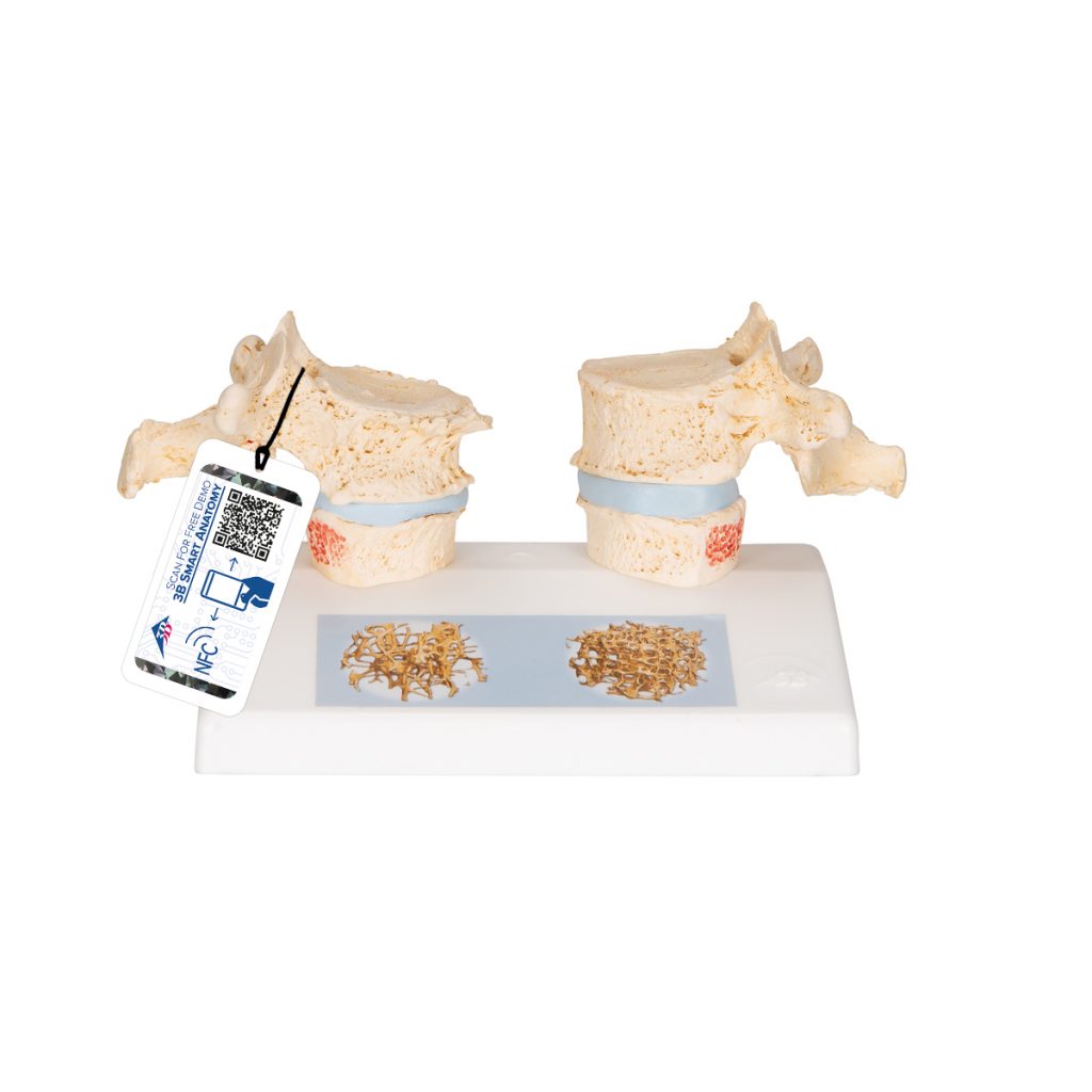
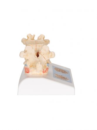
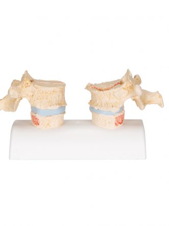
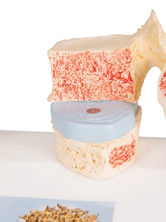
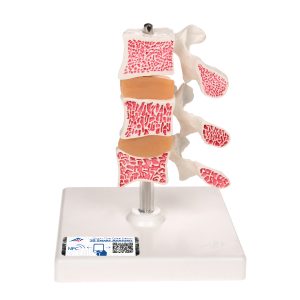
Impressive didactic model for comparing osteoporotic and normal thoracic vertebrae. Ideal for medical studies and patient consultation. The 11th and 12th thoracic vertebrae are shown.
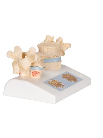
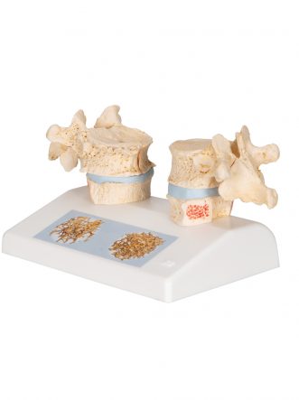
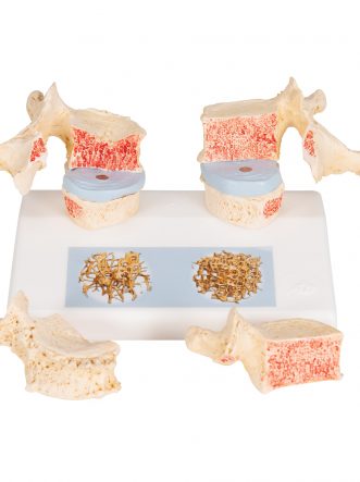
Reproductions of sequential osteoporotic thoracic vertebrae with narrower intervertebral disc are located on the left of the stand. The upper vertebra is divided in the middle. The magnetically attached vertebral half can be removed easily to show the cut surfaces. This allows clear visualization of the fractured upper part of the vertebral body caused by sintering, i.e. collapse of the bony substance in the course and as a result of osteoporosis. Degenerative changes in the bone, manifested as osteophytes, are also identifiable.
For comparison, reproductions of two corresponding healthy vertebrae with intervertebral disc are provided on the right side. One half of the upper vertebral body is magnetically attached and can be removed.
A detail illustration on the base depicts two 3D micro CT images obtained from bone biopsies. These illustrate the microacrchitecture of the osteoporotic bone, which has a lower bone density compared to healthy bone.
Osteoporosis Didactic Model — 3B Smart Anatomy
The Osteoporosis Didactic Model (Item No. 1000182 [A95], 0.238 kg) by 3B Scientific, Germany is an impressive didactic model for directly comparing osteoporotic and normal thoracic vertebrae — ideal for medical studies and patient consultation. The 11th and 12th thoracic vertebrae are shown.
Side-by-side pathological comparison in one model. Reproductions of sequential osteoporotic thoracic vertebrae with narrower intervertebral disc sit on the left of the stand, while corresponding healthy vertebrae with intervertebral disc are shown on the right — giving students and patients an immediate visual contrast rather than needing to compare two separate models.
Removable sections for internal cross-section study. The upper vertebra on each side is divided in the middle, with a magnetically attached vertebral half that can be removed easily to reveal the cut surfaces. On the osteoporotic side, this clearly shows the fractured upper part of the vertebral body caused by sintering — the collapse of bony substance that results from osteoporosis — along with degenerative changes such as osteophytes.
Micro-CT imagery grounds the model in real clinical data. A detail illustration on the base depicts two 3D micro-CT images obtained from actual bone biopsies, illustrating the microarchitecture of osteoporotic bone and its lower bone density compared to healthy bone — connecting the physical model to real diagnostic imaging.
Includes free 3B Smart Anatomy digital access. Every original 3B Scientific model includes free access to the 3B Smart Anatomy course, hosted inside the Complete Anatomy app by 3D4Medical — featuring 23 digital anatomy lectures, 117 virtual anatomy models, and 39 anatomy quizzes, plus a free warranty upgrade from 3 to 5 years upon product registration.
Why Buy from SEM Trainers? SEM Trainers & Systems is an authorised distributor of 3B Scientific products in India with over 30 years of experience supplying medical education resources to 1,500+ institutions across 28 states. We provide pan-India delivery and dedicated after-sales support.
Frequently Asked Questions
Q: Which vertebrae does this model show?
A: The 11th and 12th thoracic vertebrae, in both osteoporotic and healthy versions for direct comparison.
Q: Can the vertebrae be opened to show internal structure?
A: Yes — the upper vertebra on each side has a magnetically attached half that can be removed to reveal the cut surface and internal bone changes.
Q: Is this suitable for patient consultation, not just classroom teaching?
A: Yes — it’s specifically designed to be effective for both medical studies and explaining osteoporosis to patients directly.
Contact us at [email protected] or call +91-88495 63724 for pricing and availability.
3B Smart Anatomy explained in 90 seconds:

Reviews
There are no reviews yet.