
Description
For patient-friendly and accurate positioning learning
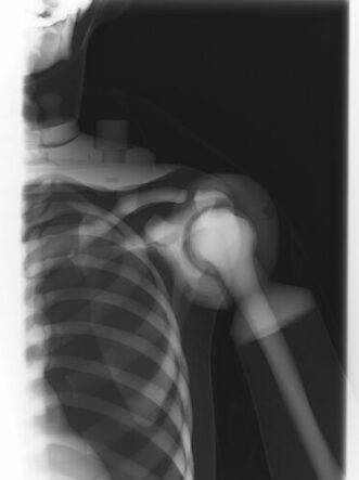
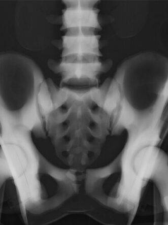
Supports scenario based trainings including communication skills
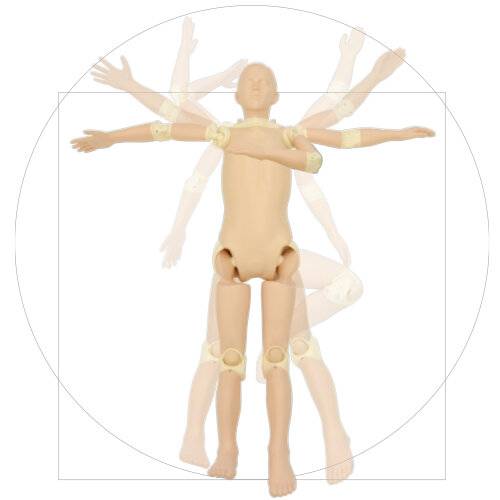
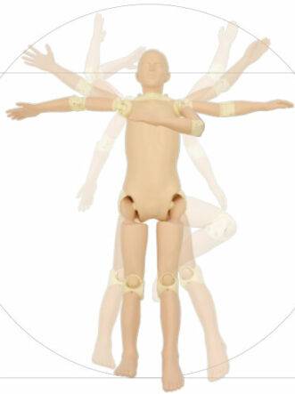
| Features | 1. Design focused on positioning and light weight with clear images. 2. Radiography images can be acquired with a lower irradiation than the real one and reduce the radiation exposure to trainees and stress on the device. 3. Each joint has the close-to-human range of motion and can be positioned according to the target part of the shot. 4. Contains all necessary landmarks for positioning 5. Specialized in positioning and has been drastically made lighter (18kg). 6. Enables training free from privacy concerns and inconveniences associated with use of standardized patients. 7. No metal parts, which causes artifacts, are included in the phantom. |
|---|---|
| Training skills / Applications | Patient positioning Patient transportation Plain radiography |
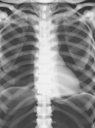
| Case / Pathology | [Skeleton] Skull, cervical spine, vertebrae, clavicles, scapulae, sternum, pelvis, lungs (without vessels), heart, kidneys, upper and lower arm bones, carpal, metacarpal, femur, kneecaps, lower leg bones, tarsi, metatarsals, phalanges.[Internal organs] trachea (up to 1st bifurcation), lungs (diaphragm only), heart, kidneys |
| Set includes | Adult whole body phantom, head stand, tools for assembly, radiography data, clothes, instruction manuals Separable into 10 parts |
| Size (approx.) | chest girth : 85cm (thickness : 20cm) waist girth : 75cm (thickness : 19cm) |
| Height (approx.) | 165 cm |
| Weight (approx.) | 18 kg |
| Materials | Soft fabrics : polyurethane foam (density 0.2) Skeleton : epoxy resin (density 1.31) Skull : urethane resin (density 1.12) |
| Production & Development Supervision | Product Supervision: Yuji Ogata, MD. Director of the Clinical Radiology Course, Faculty of Health and Medicine, Graduate School, Medical University of Morinomiya |
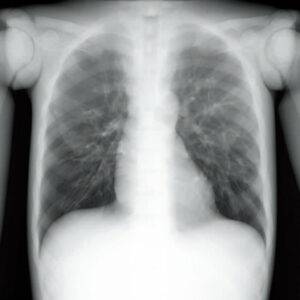
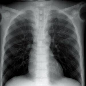
| Radiographic Condition | Because it is designed with a focus on positioning, the imaging conditions are not the same as clinical conditions for imaging of the human body. In order to reduce the operator’s radiation exposure and the stress to the device, the phantom is designed for imaging using only from one-half to one-third of the average radiation used under normal clinical conditions. |
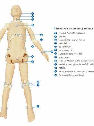
| Landmarks for positioning | External Acoustic Foramen Mastoid Seventh Cervical Vertebra Manubrium Xiphisternum Styloid Process of Radius Superior Margin of the Symphysis Pubis Medial Epicondyle of Femur/Epicondylus Lateralis Patella Malleolus (Internal condyle /External condyle) Subcostal area Landmark on the body surface Trochanter Processus styloideus ulnae |
| Update | December 2, 2020 |
Why Choose the X-Ray Training Phantom PBU-POSE for Patient Positioning Training in India?
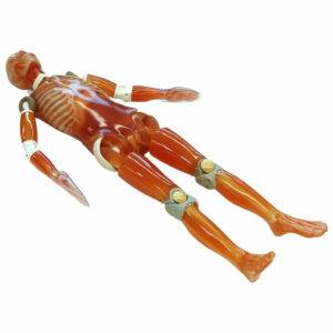
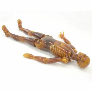
The X-Ray Training Phantom PBU-POSE (Item No. PH-79, Brand: Kyoto Kagaku, Japan) is purpose-built for patient-friendly, accurate positioning learning and supports scenario-based training including communication skills. At just 18kg, it’s dramatically lighter than standard whole body phantoms, with each joint offering close-to-human range of motion positioned according to the target imaging area. Developed under the supervision of Yuji Ogata, MD, Director of the Clinical Radiology Course, Faculty of Health and Medicine, Graduate School, Medical University of Morinomiya.
Because the phantom is designed with a focus on positioning rather than diagnostic image quality, it can be imaged using only one-half to one-third of the average clinical radiation dose — significantly reducing operator exposure and stress on imaging equipment during repeated training sessions. All necessary anatomical landmarks for accurate positioning are included, and the phantom contains no metal parts, avoiding imaging artifacts. It also enables positioning training free from the privacy concerns and inconveniences associated with using standardized (live volunteer) patients.
Training applications:
- Patient positioning
- Patient transportation
- Plain radiography
Anatomy represented:
- Skeleton: Skull, cervical spine, vertebrae, clavicles, scapulae, sternum, pelvis, lungs (without vessels), heart, kidneys, upper and lower arm bones, carpal bones, metacarpals, femur, kneecaps, lower leg bones, tarsi, metatarsals, phalanges
- Internal organs: Trachea (to first bifurcation), lungs (diaphragm only), heart, kidneys
Positioning landmarks included: External acoustic foramen, mastoid process, seventh cervical vertebra, manubrium, xiphisternum, styloid process of radius, superior margin of the symphysis pubis, medial epicondyle of femur/lateral epicondyle, patella, medial and lateral malleolus, subcostal area, trochanter, styloid process of ulna, plus additional body surface landmarks.
Set includes: Adult whole body phantom, head stand, assembly tools, radiography data, clothing, instruction manual. Separable into 10 parts.
Specifications:
- Height approx. 165cm
- Chest girth approx. 85cm (thickness 20cm); waist girth approx. 75cm (thickness 19cm)
- Weight approx. 18kg
- Materials: Soft fabrics — polyurethane foam (density 0.2); skeleton — epoxy resin (density 1.31); skull — urethane resin (density 1.12)
Why Buy from SEM Trainers? SEM Trainers & Systems is an authorized distributor of Kyoto Kagaku products in India with over 30 years of experience supplying patient positioning training equipment to 1,500+ institutions across 28 states. We provide pan-India delivery and dedicated after-sales support.
Frequently Asked Questions
Q: Can this phantom replace standardized patient training? A: It’s designed to enable positioning training free from the privacy concerns and logistical challenges of using standardized patients, though it’s typically used to complement rather than fully replace such training.
Q: How do I order from SEM Trainers? A: Contact us at [email protected] or call +91-88495 63724 for pricing and availability.
Reviews
There are no reviews yet.