

Description
New anatomy app called 3B Smart Anatomy now included for FREE with Human Female Pelvis Skeleton Model with Ligaments, Vessels, Nerves, Pelvic Floor Muscles & Organs, 6 part.
Every original 3B Scientific anatomy model now includes these additional FREE features:
- Free access to the anatomy course 3B Smart Anatomy, hosted inside the award-winning Complete Anatomy app by 3D4Medical
- The 3B Smart Anatomy course includes 23 digital anatomy lectures, 117 different virtual anatomy models and 39 anatomy quizzes to test your knowledge
- Bonus: FREE warranty upgrade from 3 to 5 years with every product registration
Human Female Pelvis Skeleton Model with Ligaments, Vessels, Nerves, Pelvic Floor Muscles & Organs, 6-part — 3B Smart Anatomy
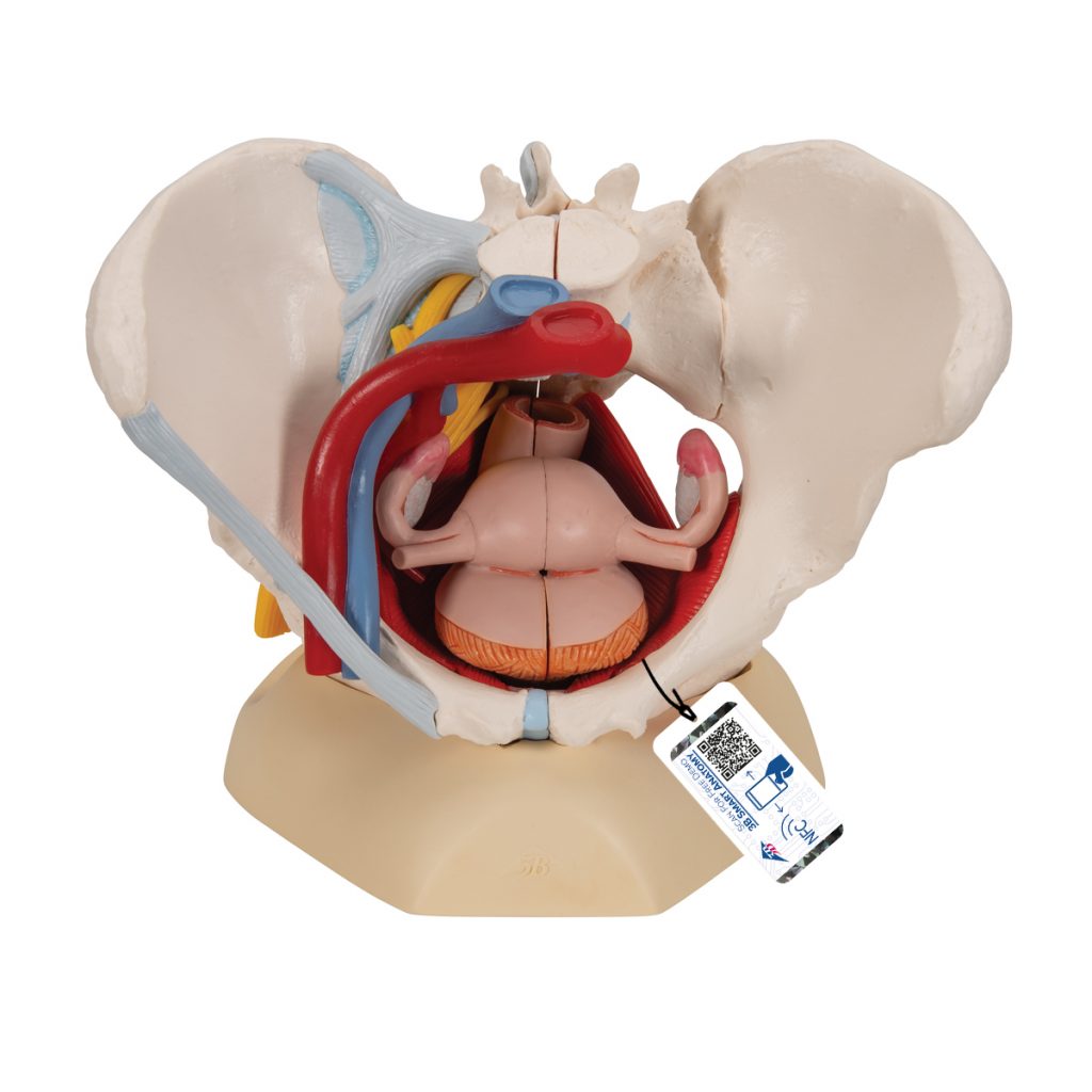
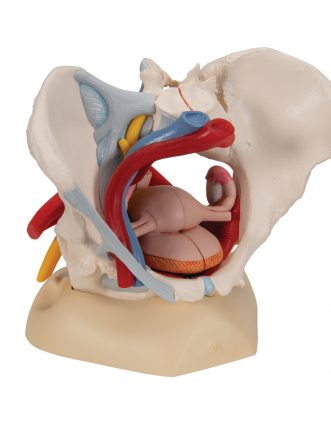
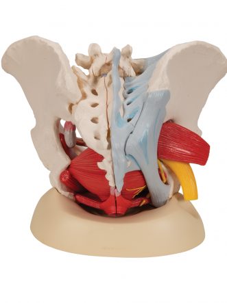
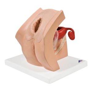
The Human Female Pelvis Skeleton Model with Ligaments, Vessels, Nerves, Pelvic Floor Muscles & Organs, 6-part (Item No. 1000288 [H20/4], 2.274 kg) by 3B Scientific, Germany presents detailed information about the topography of bones, ligaments, vessels, nerves, pelvic floor muscles, and female genital organs in a life-size six-part model.
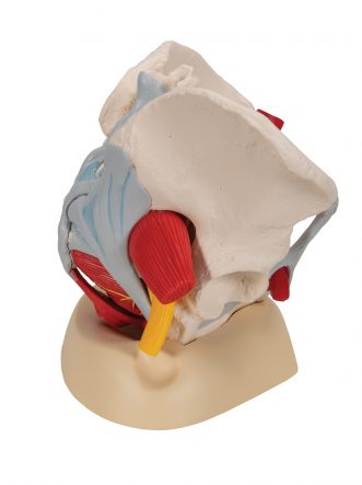
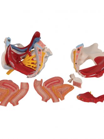
Complete pelvic floor with removable structures. The model presents the whole pelvic floor with a partially removable midsagittally sectioned external anal sphincter, external urethral sphincter, and deep and superficial transverse perineal and bulbospongiosus muscles — giving students layer-by-layer access to pelvic floor anatomy rather than a fixed, static view.
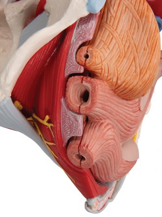
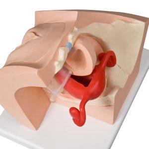
Reproductive organs that come apart for detailed study. The rectum, uterus with fallopian tubes, ovaries, and vagina are all removable and can be disassembled into both halves by midsagittal section, allowing close examination of internal structure from multiple angles.
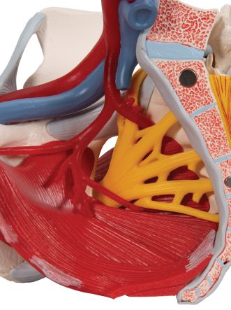
Vascular and neural anatomy shown in full detail. The right pelvic half demonstrates the divisions and topographical anatomy of the common iliac artery, external and internal iliac artery, and common iliac vein and external iliac vein, along with the right sacral plexus, right sciatic nerve, and right pudendal nerve.
Skeletal structure with genuine articulation. Bones and ligaments presented include two hip bones, the pubic symphysis, the sacrum and coccyx, and the fifth lumbar vertebra with intervertebral disc. A midsagittal section through the fifth lumbar vertebra, sacrum, and coccyx allows both pelvic halves to be disassembled, revealing part of the cauda equina in the vertebral canal, with the left half of the fifth lumbar vertebral body also removable. The right half additionally shows the inguinal ligament, sacrotuberous ligament, sacrospinous ligament, anterior sacroiliac ligaments, iliolumbar ligament, anterior longitudinal ligament, interosseous sacroiliac ligament, posterior sacroiliac ligament, and obturator.
Ideal for detailed study of female genital and pelvic anatomy in gynecology, obstetrics, and general anatomy education.
Includes free 3B Smart Anatomy digital access. Every original 3B Scientific model includes free access to the 3B Smart Anatomy course, hosted inside the Complete Anatomy app by 3D4Medical — featuring 23 digital anatomy lectures, 117 virtual anatomy models, and 39 anatomy quizzes, plus a free warranty upgrade from 3 to 5 years upon product registration.
Why Buy from SEM Trainers? SEM Trainers & Systems is an authorised distributor of 3B Scientific products in India with over 30 years of experience supplying medical education resources to 1,500+ institutions across 28 states. We provide pan-India delivery and dedicated after-sales support.
Frequently Asked Questions
Q: What anatomical systems does this model cover?
A: Pelvic bones and ligaments, the pelvic floor muscles, female reproductive organs, and the associated vessels and nerves — a comprehensive 6-part model.
Q: Can the reproductive organs be removed for closer study?
A: Yes — the rectum, uterus with fallopian tubes, ovaries, and vagina are all removable and can be disassembled by midsagittal section.
Contact us at [email protected] or call +91-88495 63724 for pricing and availability.
TIP: You will also receive access to a free 3-day trial to all premium features of the Complete Anatomy app when you sign up for your 3B Smart Anatomy course.
To unlock these benefits, simply scan the label located on your model and register online. All 3B Smart Anatomy features are completely free of charge for you. Click here to learn more.
The rectum, uterus with fallopian tubes, ovaries and vagina are also removable and can be disassembled into both halves by midsagital section. The right pelvic half demonstrates the divisions and topographical anatomy of the common iliac artery, the external and internal artery and also of the common iliac vein and the external iliac vein. The right sacral plexus, right sciatic nerve and right pudendal nerve are also shown.
Bones and ligaments presented: Two hip bones, the pubic symphysis, the sacrum and the coccyx, the fifth lumbar vertebra with intervertebral disc. A midsagital section through the fifth lumbar vertebra, sacrum and coccyx, allow both halves of the pelvis to be disassembled revealing a part of the cauda equina in the vertebral canal. The left half of the fifth lumbar vertebral body is removable. The right half of the model shows the following pelvic ligaments: inguinal ligament, sacrotuberous ligament, sacrospinous ligament, anterior sacroiliac ligaments, iliolumbar ligament, anterior longitudinal ligament, interosseous sacroiliac ligament, posterior sacroiliac ligament and obturator.
This model is great for detailed study of the female genital and pelvic anatomy.
3B Smart Anatomy explained in 90 seconds:


Reviews
There are no reviews yet.